“At Family and Cosmetic Dentistry of Kokomo, we take pride in being the best dentists in Kokomo. Our philosophy centers around a patient-centered approach, where empathy and compassion are paramount. We aim to create a warm and welcoming environment, making patients of all ages feel comfortable during their visits. By investing in cutting-edge technology, we offer a comprehensive range of services, ensuring efficient and effective treatments.
Patient education is essential to us, empowering individuals to make informed decisions about their oral health. We prioritize building long-term relationships and are committed to providing personalized care for new and returning patients to help our patients achieve optimal oral health and maintain beautiful smiles for life.
Sure we’re dentists. But we’re proud Americans and nice folks just like you, too. We know too many dentists feel old-fashioned, out-of-touch, and intimidating. Our team at Family and Cosmetic Dentistry of Kokomo is changing all that. See for yourself why our dentist office is the place to go for friendly, modern, and comfortable dental care for all ages. Our goal is to make your visit as comfortable as possible.”
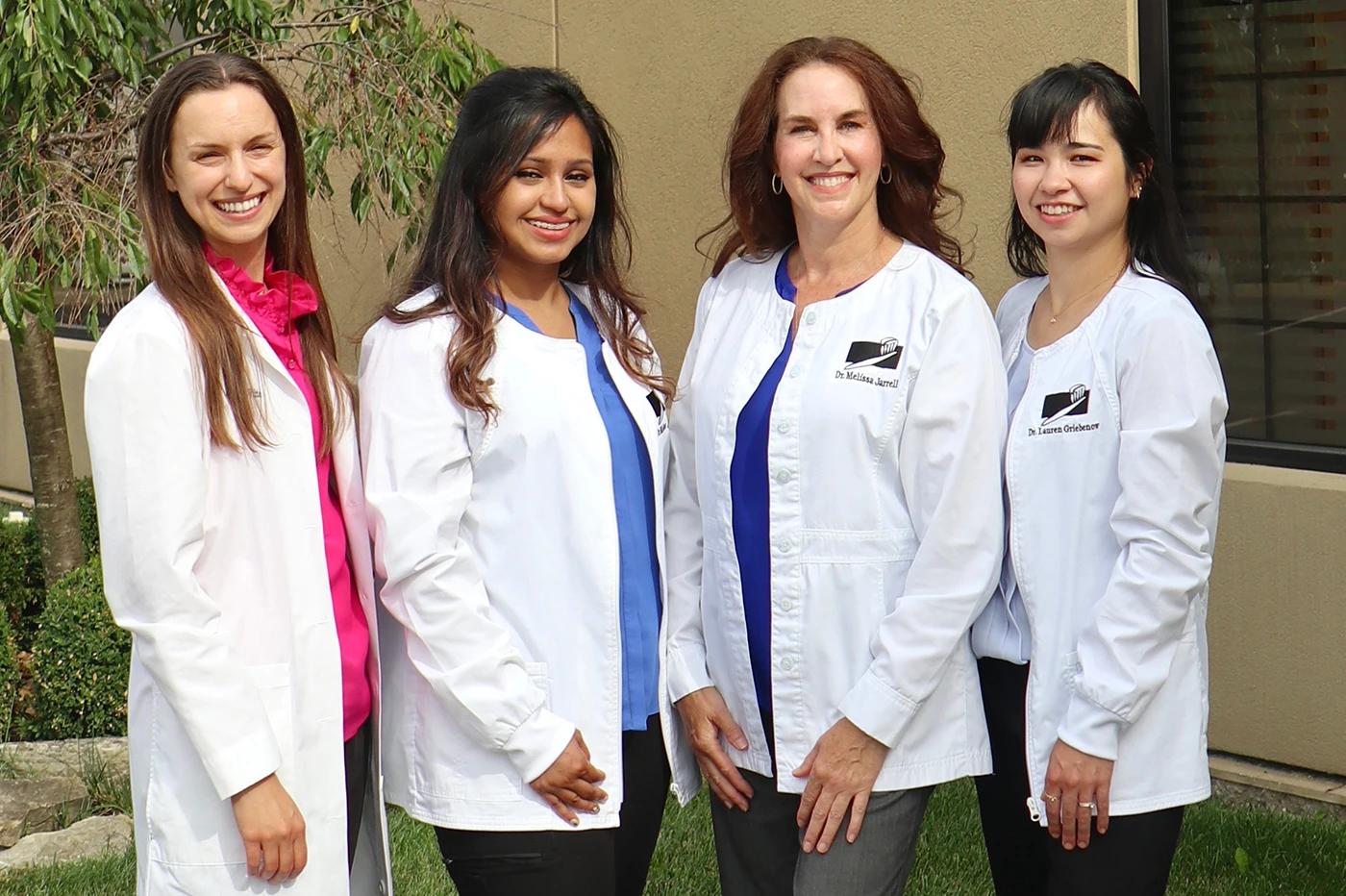
-Dr. Melissa Jarrell, Dr. Lauren Griebenow, Dr. Hannah White, Dr. Tamanna Azim
Dr. Melissa Jarrell
Dr. Lauren Griebenow
Dr. Hannah White
Dr. Tamanna Azim
Dr. Melissa Jarrell and team have been serving the dental needs of their Kokomo patients with personal attention and quality professional care for over 25 years.
Our goal at Family and Cosmetic Dentistry of Kokomo is to provide compassionate high quality dental services using the latest techniques and allowing you to have healthy teeth and a beautiful smile for a lifetime!
View our comfort features
- Refreshment Bar: chilled bottled water and juice
- iPad in each private room
- Free Wi-Fi
- Music on our iPad or your own personal device with headphones
- Tylenol/Motrin before or after treatment
- Protective Tinted Eye-wear
- Lip Balm
- Too cool? Warm, Comfy Blankets
- Too warm? Personal fan
- Supporting Neck Pillow
- Knee Pillows: For extra support
- Mouth Prop to rest your jaw during procedures
- Nitrous Oxide Sedation “laughing gas”
- Sedative “sleeping” pill (Oral Sedation)
- IV “twilight” Sedation
- Payment plans (finance options available)
- Warm moist towel “to refresh” after dental treatment
Add these comfort features to the most advanced equipment and techniques available today to get an unsurpassed dental experience and result.
Can a dentist really make you smile? You bet!
Meet the Dentists
We welcome the opportunity to care for your dental needs and will do everything possible to make your visits pleasant.
Learn More

Smiles for Vets
Spreading FREE Smiles Across Howard County Indiana
Free Dental Care for Veterans on
Friday Sept. 11, 2026
Learn More

Same Day Crowns
Dr. Melissa A. Jarrell and team are certified to use the CEREC® crown restoration system enabling patients to receive a custom fitting crown in an hour.
Learn More

Sedation Dentistry

“Do you take my insurance?”
IT’S THE QUESTION ON EVERYONE’S MIND
Whether you have dental insurance or not, our doors are open to you. We’re in-network with Delta Dental (PPO and Premier). Although, we are not in network with other PPO
insurances we will file your insurance and help you get the most out of your dental
benefits. If you’re not insured, our dental office offers flexible in-office financing (Preventive Care Plan) and payment options that can fit any budget.
At this time, we DO NOT ACCEPT MEDICAID OR NON-PPO MEDICARE.
We apologize for the inconvenience.

Why People Love Family and Cosmetic Dentistry of Kokomo?
Posted on